

Extremity PCCT
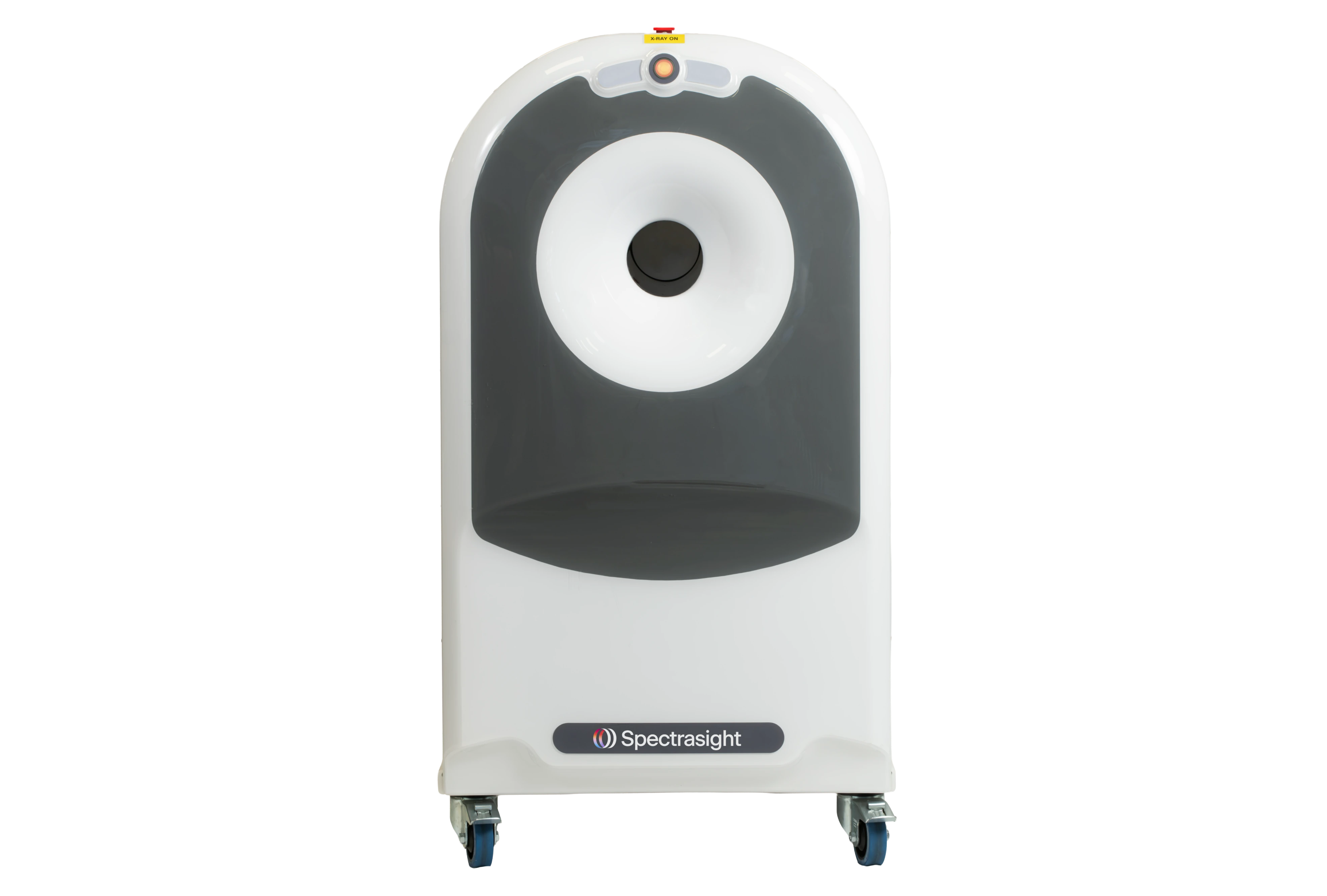
Designed exclusively to support hand and wrist specialists. Premium photon counting CT imaging, where your patients are, from the start to the end of the pathway.
Ultra-High Resolution at Ultra-low dose
Ultra-High Resolution (UHR) of 33 lp/cm with 0.09mm isotropic voxels¹ with the Standard protocol. Sharp bone and soft tissue detail, with native metal artifact reduction² . Clearly visualize fine trabecular detail and subtle cortical breaks. 5x lower dose³ plus integrated shielding⁴.
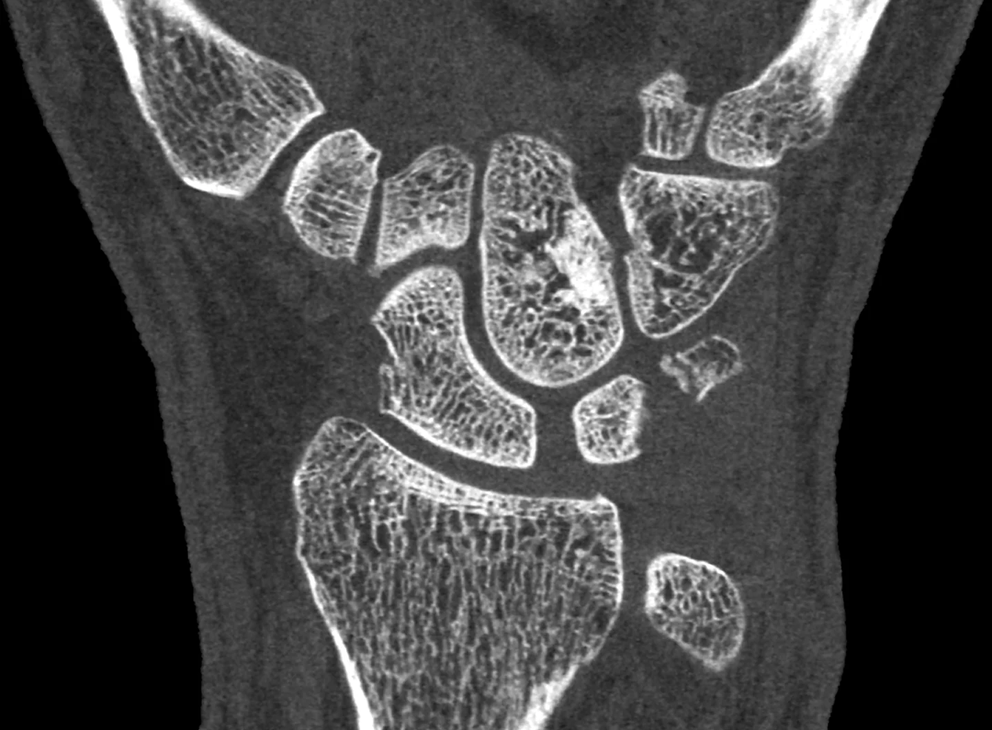
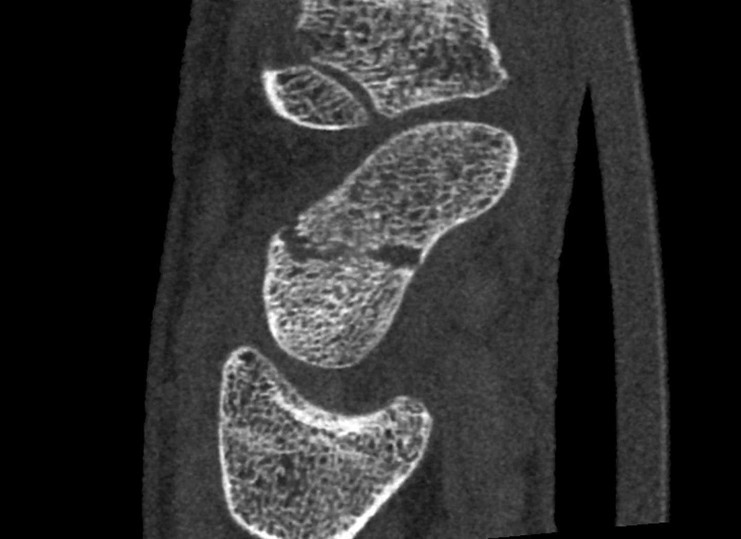
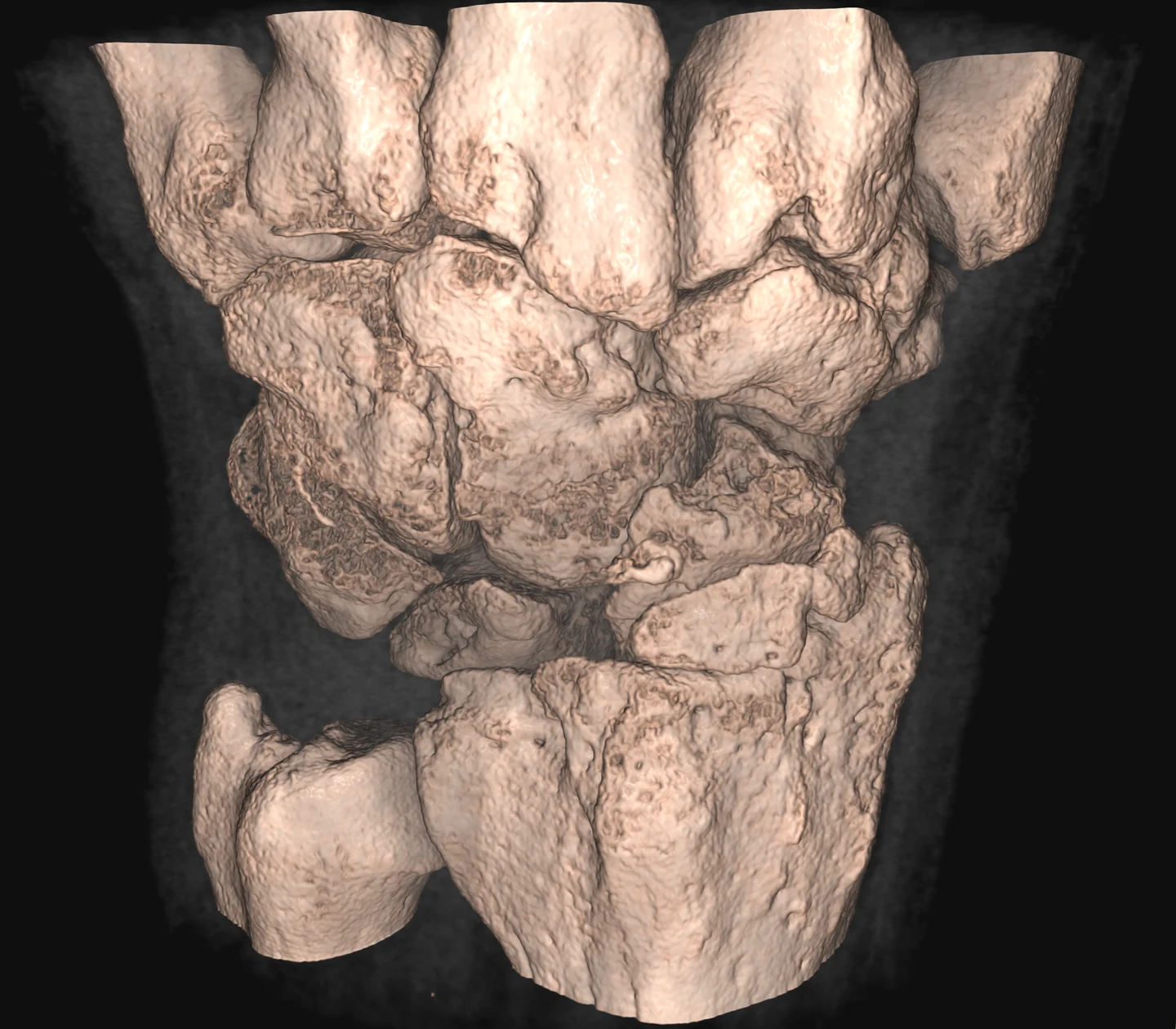
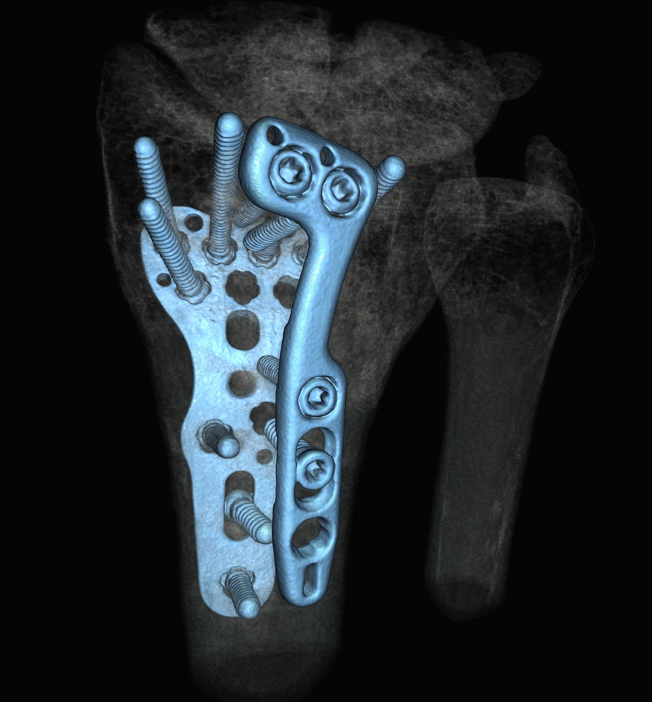
Scan now, see clearly.
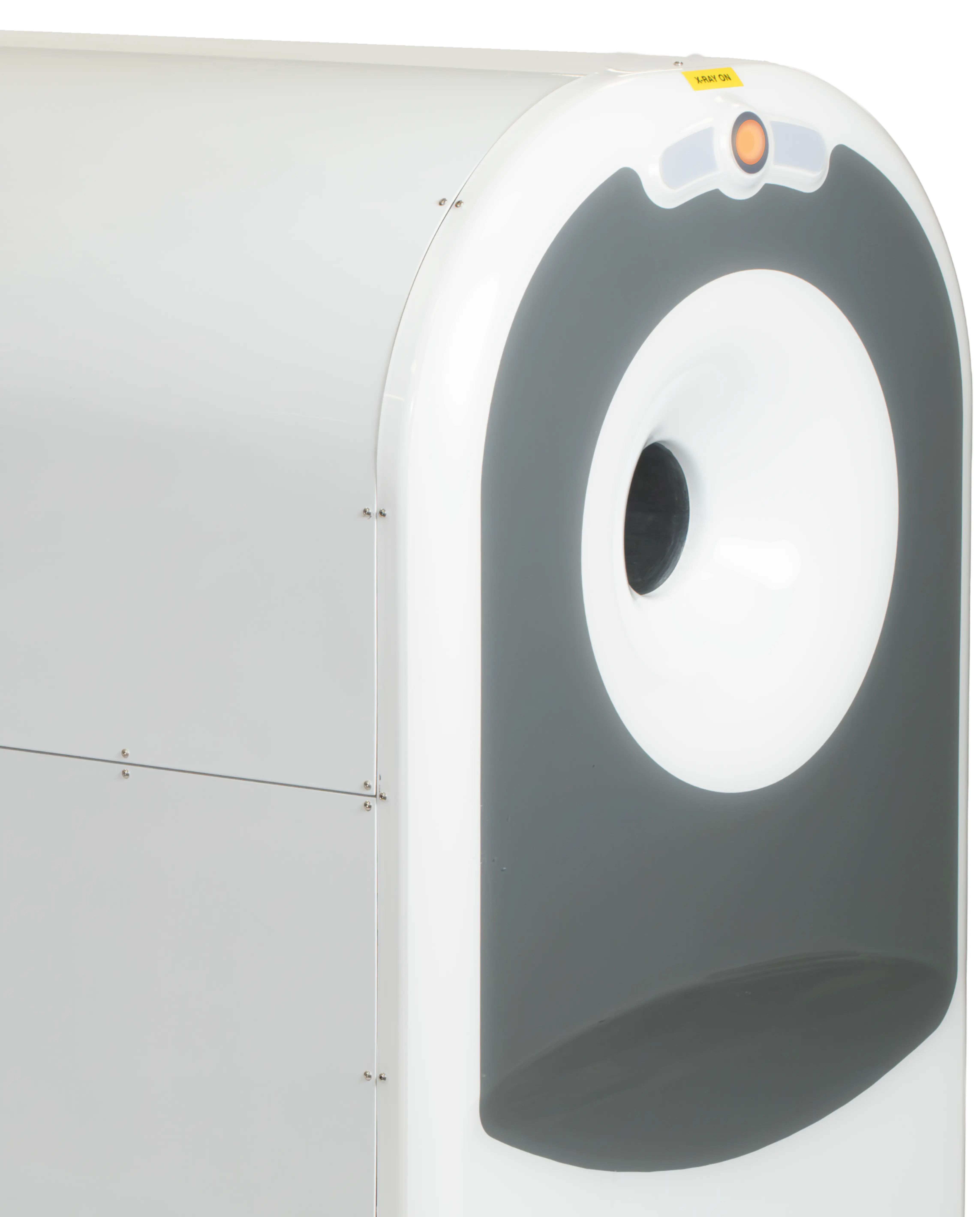
Quick plug’n’play installation and commissioning within existing infrastructure. Simple protocols with DICOM/PACS integration. Small footprint is easy to move between facilities and fits into smaller sized rooms.
ED & Urgent Care
Image fractures at the front door instead of moving through to radiology
Clinic & Office
Specialist clinics, ASCs, orthopedic, sports medicine and radiology at standard exam room scale
Relocatable
Extremity PCCT can be moved between sites with minimal supporting infrastructure changes
High-tech imaging, made simple.
Photon-counting detectors count individual X-ray photons directly instead of measuring light from a scintillator. Spectral imaging inherently handles electronic noise allowing clear visualization of both anatomical and metallic structures. The system uses a Cadmium-Telluride (CdTe) detector based on Medipix3 technology.
Technical specifications
Detector
CdTe PCD
X-ray Tube
25 KHU
Tube Current
0.010 – 0.35 mA
Bore
120 mm
Footprint⁵
0.8 m² (8.6 ft²)
Weight
405 kg (890 lb)
Software
MARS Scanner, MARS Vision
Frequently asked questions
It is a photon-counting CT system cleared by the FDA for x-ray computed tomography imaging of upper extremities in adult patients, for use by qualified and trained healthcare professionals (510(k) K252249). Prescription use only.
Photon-counting detectors count individual X-ray photons directly instead of measuring light from a scintillator, which largely excludes electronic noise and allows smaller detector elements. The system uses a CdTe detector based on Medipix3 technology. Energy-resolved acquisition is inherent to photon counting; we make no material-differentiation claims for this system.
Ultra-High Resolution (UHR) of 33 lp/cm with 0.09 mm isotropic voxels. That is measured on the standard wrist protocol with bone reconstruction, not a special high-dose mode.
5 times lower dose.3 The tube operates at 0.010–0.35 mA, and dose for any given examination depends on the protocol selected.
The gantry bore is 12 cm (4 ¾ inch) and the patient stays seated with the limb positioned into it. There is no table and no open tunnel. The maximum scan length is 44 cm (17 ¼ inch). In practice this covers the fingers, hand, wrist and forearm.
The scanner occupies about 0.8 m², is mounted on wheels and runs on standard mains power. The device is internally shielded and may not need a purpose-built lead-lined room, but shielding, machine registration and survey requirements are set by your state radiation control programme, so confirm the position locally before installation.
The cleared indication requires operation in a professional healthcare environment by qualified and trained healthcare professionals. Operator credentialing for CT is set by state law, so confirm the requirement in your state; interpretation remains with a suitably credentialed physician.
Images can be acquired with metalwork in the field of view. Blooming or beam hardening is minimized, increasing the clarity of bone-metal interfaces. Combined with a UHR output of 0.09 mm isotropic voxels, even small details like screw threads can be visualized in the images.
Images are acquired and reviewed in MARS Vision, our post-processing software, which provides multiplanar reformats and 3D rendering. The system is configurable for integration with DICOM/PACS export protocols.
CT imaging of the upper extremity is described in the USA by CPT codes 73200–73202. Coverage, accreditation requirements and payment depend on your payer and your setting and we do not guarantee payment. Contact us for the technical documentation your billing team will need. Non-US countries will have different coding systems and requirements.
See Spectrasight in action.
Book a demonstration with our clinical
applications specialists
2. Metal artifact reduction is achieved through features native to the technology. The system does not include MAR algorithms.
3. Five times lower dose measured in a manufacturer study of CTDIvol for recommended wrist protocols on the MARS Extremity vs a 64-slice MDCT, using a 10 cm CTDI dosimetry phantom.
4. Lead-shielded room may not be required, subject to applicable State and site radiation control requirements.
5. Scanner footprint only; excludes workstation and operator space. System is mobile but must be operated stationary.
